The electron microscope stands as a pinnacle of scientific achievement, a technological wonder that has revolutionized our understanding of the microscopic world.
With its unparalleled ability to reveal intricate details at the atomic and molecular level, this extraordinary instrument has paved the way for countless breakthroughs in various scientific disciplines.
From biology to materials science, the electron microscope has become an indispensable tool for researchers seeking to unlock the secrets of the unseen.
Unlike its optical counterpart, which uses visible light to magnify objects, the electron microscope employs a beam of electrons to illuminate and visualize specimens.
The use of electrons, with their shorter wavelengths, enables a level of resolution unattainable by traditional light microscopes.
It allows scientists to observe structures as small as a few picometers, unveiling a realm of intricate details that were once hidden from view.
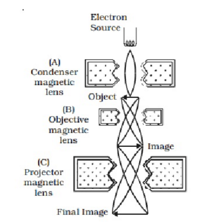
The working principle of the electron microscope involves several key components. First, a source, typically a heated filament or a field emission gun, generates a beam of electrons.
The beam is then accelerated by an electric field towards the specimen under investigation. In transmission electron microscopy (TEM), the electron beam passes through the specimen, and a series of magnetic lenses focus the electrons to form an image on a fluorescent screen or a digital detector.
In scanning electron microscopy (SEM), the beam is scanned across the surface of the specimen, and the emitted secondary electrons or backscattered electrons are detected to create an image.
Types Of Electron Microscopes:
1) Scanning Electron Microscope (SEM)
2) Transmission Electron Microscope (TEM)
Advantages of Electron Microscopy:
1) High Resolution:
Electron microscopes offer unparalleled resolution, enabling scientists to visualize objects at the atomic and molecular level. This high resolution is crucial for studying the intricate details of biological structures, materials, and nanoparticles.
2) Magnification:
Electron microscopes can achieve magnifications ranging from thousands to millions of times, providing a detailed view of the specimen. This level of magnification is particularly useful in fields such as nanotechnology and materials science.
3) Versatility:
Electron microscopy encompasses various techniques and modalities, including TEM, SEM, and cryo-EM. Each technique offers unique advantages, allowing researchers to tailor their approach to specific research questions and sample types.
4) Sample Flexibility:
Electron microscopes can analyze a wide range of specimens, including biological samples, materials, cells, tissues, and nanoparticles. With appropriate sample preparation techniques, electron microscopy can provide insights into the structure, composition, and morphology of diverse materials.
5) Real-time Imaging:
Electron microscopes can capture images and videos in real-time, allowing researchers to observe dynamic processes and interactions at the nanoscale. This capability is vital for understanding biological mechanisms and studying materials under various conditions.
Disadvantages of Electron Microscopy:
1) Complex Sample Preparation:
Preparing samples for electron microscopy often requires specialized techniques, such as fixation, dehydration, staining, or cryo-preservation. These processes can be time-consuming and may introduce artifacts that affect the specimen's natural state.
2) High Cost:
Electron microscopes are sophisticated instruments that come with a high price tag. The initial investment, as well as the maintenance and operational costs, can be a significant barrier for many research institutions and laboratories.
3) Limited Sample Size:
Electron microscopes typically have a small sample chamber, restricting the size of specimens that can be analyzed. In some cases, samples need to be sectioned or prepared as thin films, which may alter their properties.
4) Vacuum Requirements:
Electron microscopy requires a vacuum environment to ensure that the electron beam is not scattered or absorbed by air molecules. This constraint limits the ability to examine live or hydrated samples directly.
Applications of Electron Microscopy:
Certainly! Electron microscopy finds applications in a wide range of scientific disciplines. Here are some additional areas where electron microscopy is extensively used:
1) Biological Research:
Electron microscopy plays a crucial role in biological resaerch
2) Materials Science:
Electron microscopy is indispensable in materials science research. It enables the characterization of materials at the atomic and nanoscale level, providing insights into crystal structures, defects, and interfaces. This information is vital for designing new materials with improved properties and understanding the behavior of materials under different conditions.
3) Nanotechnology:
With its ability to visualize nanoscale structures, electron microscopy is a fundamental tool in nanotechnology. It helps researchers examine nanoparticles, nanowires, and nanodevices, enabling the precise characterization of their size, shape, and composition. This knowledge aids in the development of advanced nanomaterials and nanotechnologies for various applications, including electronics, energy storage, and biomedical devices.
4) Forensic Science:
Electron microscopy plays a crucial role in forensic investigations. It assists forensic scientists in analyzing trace evidence, such as fibers, paint chips, or gunshot residue, to identify sources and establish connections. Electron microscopy can provide detailed information about the morphology, composition, and elemental analysis of these materials, aiding in criminal investigations.
5) Geology and Earth Sciences:
Geologists and earth scientists use electron microscopy to study rocks, minerals, and geological samples. It allows for the examination of mineral compositions, texture, and crystal structures, aiding in the identification of minerals and the understanding of geological processes. Electron microscopy also helps in the analysis of meteorites and extraterrestrial materials, providing insights into the formation and evolution of celestial bodies.
6) Pharmaceutical Research:
Electron microscopy is employed in pharmaceutical research and development. It assists in the characterization of drug particles, including their size, shape, and surface properties. Understanding the morphology and structure of pharmaceutical compounds is crucial for drug formulation, delivery systems, and quality control.
7) Environmental Science:
Electron microscopy is utilized in environmental science to study pollutants, particulate matter, and contaminants. It aids in the analysis of aerosols, microplastics, and airborne particles, providing insights into their sources, composition, and potential environmental impact. Electron microscopy is also valuable in studying soil and water samples, helping to identify pollutants and understand their interactions with the environment.
8) Archaeology and Cultural Heritage:
Electron microscopy plays a significant role in the analysis and preservation of artifacts and cultural heritage. It enables researchers to examine ancient materials, pigments, and coatings, assisting in the identification of materials used, degradation mechanisms, and restoration efforts. Electron microscopy is particularly useful in the study of delicate and valuable objects where non-destructive analysis is required.
9) Electronics and Semiconductor Industry:
Electron microscopy is essential for the semiconductor industry. It aids in the development and quality control of electronic devices by examining semiconductor materials, integrated circuits, and nanoscale features. Electron microscopy enables the visualization of device structures, identifying defects, and optimizing manufacturing processes.
These applications represent just a fraction of the diverse fields where electron microscopy finds utility. The remarkable capabilities of this technology continue to drive scientific advancements and contribute to our understanding of the world at the smallest scales.
 |
| Image Under Electron Microscope. |


0 Comments